
Grand Jury
November 13, 1974
Testimony of Dr. Frank Gemma, M.D.
Scans of original transcript


I, Mary M. Ritchie, being a Notary Public in and for the State of North Carolina, was appointed to take the testimony of the following witness, Frank Eugene Gemma, before the Grand Jury, Raleigh, North Carolina, commencing at 11:15 a.m. on November 13, 1974. All Grand Jurors present.
Whereupon, FRANK EUGENE GEMMA, having been first duly sworn, was examined and testified as follows:
EXAMINATION BY MR. WOERHEIDE:
Q Will you state your full name, please, sir?
A Frank Eugene Gemma.
Q And are you a medical doctor?
A Yes, sir.
Q What's your home address?
A My home address is 104A Scoffield Circle, Ft. Riley, Kansas.
Q And are you in the armed forces?
A Yes. I am.
Q What's your military title and your post of duty, sir.
A My military title is Chief, Department of Surgery, Erwin Army Hospital.
Q And, directing your attention to February, 1970, were you -- what was your position?
A At that time I was Chief of the General Surgery Service at Womack Army Hospital.
Q All right, sir. And that's at Fort Bragg, North Carolina. Is that correct?
A That's correct.
Q Now, directing your attention to the morning of February 17, 1970, do you recall being summoned to examine and give medical care and attention or surgical procedures to one Captain Jeffrey MacDonald?
A Yes. I do.
Q Approximately what time was that, sir?
A It was sometime between six-thirty and seven o'clock in the morning.
Q Now, where did you first meet and observe Captain MacDonald?
A He was on the intensive care ward on the second floor of the hospital.
Q Well, tell us about his appearance, what you observed so far as his -- any physical injuries that he might have, what you observed so far as his behavior and conduct were concerned.
A At the time that I first saw him he had been given medication by one or two of the other physicians that had seen him before, so he was somewhat sedated, but at the same time both a little bewildered and anxious would be the best way that I could describe his general appearance. He appeared and was asking questions as far as how his family was, what had happened to them and is everything going to be all right? To the best of my recollection these are the only concern [sic] that he really had at that particular time.
The doctor that called me in was concerned as to whether the pneumothorax that he had was significant enough to require a chest tube.
Q Who was the doctor that called you in?
A Dr. Bronstein.
Q Do you know Dr. Jacobson?
A Yes, sir.
Q Was he also in attendance that morning?
A Yes, he was.
Q And do you know Dr. Straub?
A I should know Dr. Straub, and, if I see him I'm sure I would recall him, but I really can't remember him.
Q Well, he's a radiologist. Do you remember a radiologist being there that morning?
A No. I don't remember seeing a radiologist that morning.
Q Now, tell us a little bit more, if you can, what he was saying. Did he recount in any way what had happened early that morning as far as he was concerned so far as his family?
A No. I don't remember. In fact, I'd probably made a conscientious effort to avoid discussing this with him. Dr. Bronstein had briefed me on the way to the intensive care ward as to what had happened to his knowledge and that Dr. MacDonald did not know that his wife and children were, in fact, dead. So the question in our minds were when to tell him definitely that they were no longer alive. So we tried to concentrate on his condition rather than get involved with how his family was.
Q You were concerned with his physical condition and giving him whatever medical and surgical treatment that he might require. Is that it?
A And, if he asked -- I'm not positive, but he
Webmaster note:
Page 6 missing from copy of document received. Page 7 continues below.
Q All right, sir. I show you a couple of diagrams that have been marked as Gemma exhibits one and two respectively. Exhibit #1 consisting of a male figure, seen from the front and the rear. And #2 consisting of figures of the head showing a front view and side views. Directing your attention to this point where I see the notation "stab wound, sixth or seventh I.C.S." Would that be the approximate location of the stab wound you saw, sir?
A Yes, sir.
Q And with respect to the figure from the rear it states "no stab wounds and no injury recalled on back." Does that reflect your observation?
A That is correct.
Q With regard to exhibit two, I see the full face, frontal view of the head, abrasion and small hematoma. Does that indicate the location of the hematoma that you observed, sir?
A Yes, sir.
Q Now, you listened to his chest sounds, of course, in the examination you made.
A Yes, sir.
Q And what did you observe?
A The breath sounds over the right side of the chest were diminished compared with what you heard on the left side of the chest.
Q And what did that indicate?
A This would indicate that not as much air was being taken into the lung on the right side as on the left.
Q And that tended to confirm the diagnosis previously made of a pneumothorax condition. Is that correct?
A Yes.
Q Now, were x-rays taken?
A Yes. X-rays had been taken at that time.
Q They confirmed this condition?
A Yes. They did.
Q I have a series of x-rays here. And to the extent that you think it might be useful to illustrate your testimony, I'm going to ask you to make use of these x-rays and demonstrate to the jurors what you observed and what conclusions you arrived at as a result of these observations. You can step down here, Dr. Gemma, and --
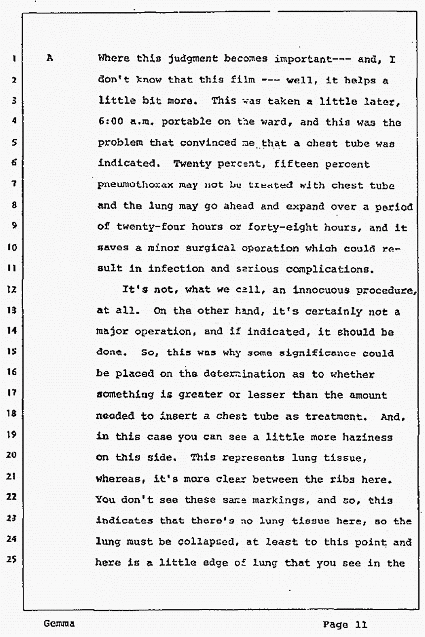
A This is the first x-ray that was taken, probably in the emergency room. And, unless, even a radiologist or someone, looked very closely, they might think this was perfectly normal. It was taken in inspiration, but it's probably a pneumothorax here, but it's very hard to tell. For that reason, when it's equivocal, especially, we take a film with the patient exhaling and when that happens it's a little bit easier. This is with no attempt to have the air fill -- the lungs filled with air. If there is air in the pleural space the lung will fall away from the chest wall more and you can see the edge of it. And this, then, this area in here is what is termed as a pneumothorax.
There is discrepancy among physicians as to estimation of the size. The initial observation either on this film, or the first film, or maybe the third one, Dr. Straub, being a radiologist, called it twenty percent. Whether that has anything to do with whether surgeons exaggerate or not I estimated this as forty percent pneumothorax, because you have to imagine that if the lung were completely out of here that would be -- or completely collapsed, and if the lung collapsed it would only collapse down to a little size, maybe a little less than your fist here. And that would be a hundred percent collapse or a hundred percent pneumothorax. So, if you take the area around here where this has fallen away and imagine the rest of this area with the exception of what would be a hundred percent, then you can say this may represent as much as forty or fifty percent of what this lung could collapse all the way.
However, in looking superficially at this, and saying, "Well, this looks to me like this is maybe only fifteen or twenty percent of the total area," neglecting the volume that the lung has to occupy when it's completely collapsed. So, that accounts for some variability, one physician calling it twenty percent and one person saying that it's forty percent and that's really a minor difference.
Q In any event, these figures are not precise. They are approximate. Is that it?
A Right.
Q It's a matter of judgment.
A Where this judgment becomes important -- and, I don't know that this film -- well, it helps a little bit more. This was taken a little later, 6:00 a.m. portable on the ward, and this was the problem that convinced me that a chest tube was indicated. Twenty percent, fifteen percent pneumothorax may not be treated with chest tube and the lung may go ahead and expand over a period of twenty-four hours or forty-eight hours, and it saves a minor surgical operation which could result in infection and serious complications.
It's not, what we call, an innocuous procedure, at all. On the other hand, it's certainly not a major operation, and if indicated, it should be done. So, this was why some significance could be placed on the determination as to whether something is greater or lesser than the amount needed to insert a chest tube as treatment. And, in this case you can see a little more haziness on this side. This represents lung tissue, whereas, it's more clear between the ribs here. You don't see these same markings, and so, this indicates that there's no lung tissue here, so the lung must be collapsed at least to this point and here is a little edge of lung that you see in the lower lobe almost completely collapsed. This line here represents the bone and the scapular. So it's not -- that isn't the line of the pneumothorax although it corresponds pretty well to where it is in that case.
Q Dr. Gemma, you testified as a witness at the Article 32 hearing, didn't you?
A No. I don't think I testified at all at that.
Q Well, in reading over some of the testimony in the Article 32 hearing the question was raised concerning a possible tension pneumothorax. Can you tell us what a tension pneumothorax is and whether or not Captain MacDonald, whether there was any indication that he had a tension pneumothorax?
A There was never any indication that he had a tension pneumothorax. A tension pneumothorax refers to this condition -- this pneumothorax as air outside the lungs but in the chest which is trapped there. It increases in size, volume, so that it can -- not only compresses the lungs on that side of the chest but actually will push the heart to the other side. And, when this happens, over a very short period of time, it puts a twist or tension on the arteries and veins coming out of the heart, affectively shutting that off and can cause death in a very short period of time. The only way that this can take place is for a ball valvelike mechanism to exist. That is, the hole in the surface of the lung has to allow air to escape out of the lung into this closed chest space, but not back through that hole into the lung and out the bronchial passages. So, anytime there is a stab wound, a hole from outside of the chest, there is relatively little possibility of a tension pneumothorax. You can't say unequivocally because there could be the possibility of such a ball valve mechanism acting and this puncture wound being filled enough that it would not allow air to escape. However, something as large as a small stab wound would almost always allow air to escape and not build up such a tension to push the heart to the other side.
Q While we are on this subject, what is the standard procedure if there is a tension pneumothorax?
A The standard procedure is to relieve it by insertion of a chest tube, but if it's an emergency situation you can relieve it with even a needle. Anything that will let the air to the outside because there is more air pressure within the chest than there is outside under that condition.
Q Once again, there is no indication whatsoever of a possible tension pneumothorax on the part of Captain MacDonald?
A No. There was never any question in my mind that that could exist.
Q All right, sir. Now, can you tell us after you determined that the chest tube was indicated, by reference to the x-rays, the diagrams that are on the wall, and I have here the testimony record of Captain MacDonald's, and I also have some photographs indicating scars that resulted from surgical procedures and also the scar of the injury which caused the pneumothorax. Can you just tell the Grand Jury in your own way what you did and what happened? Also, I have here, which may help to illustrate your testimony, surgical devices and two tubes of different shapes.
A This tube was the best that was available to represent the type of tube that was used to decompress the lung. This, however, is used for another purpose. This has two, this is two tubes, one within another tube, to take fluid from a closed space like the abdominal cavity under the skin, but not from the chest under ordinary circumstances. This tube, however, was just a little bit larger than this and looked almost exactly like it, if you can imagine the whole tube having the consistency of just this end of it. It has a radiopague on it that helped determine where it is located in the chest.
Now, since Dr. MacDonald was stabbed, there was always the possibility of blood accumulating in the chest, not rapidly enough to cause shock, because we know from his pulse and his blood pressure that he was not in clinical shock. However, you can lose as much as a pint of blood that would not even show up. This sort of gets in this -- spreads itself out over the diaphragm in such a way that it isn't really apparent on x-ray. This doesn't have to be blood. It could be fluid from some other disease that can accumulate up to about 500 cc's or a pint. However, any more than that would usually start giving some haziness and if there is air in the chest, too, there will be a straight line where the air and the fluid meet.
Whereas, if there's no air, no pneumothorax, and you have bleeding or fluid in the chest, it's less apparent because it will push the lung away all along the surface and there's a pretty big surface area of the lung. And, there's also three different lobes so there is [sic] spaces it can get in between and until the fluid gets to be close to two pints, even then it might be missed without air. With air in the chest with a hemothorax anything over a pint we would be able to see. But, even that's important to get out of the chest if it exists there. It's a source for promoting infection and so we want to get rid of it.
So, for that reason with a wound causing the pneumothorax, we usually put a tube in the lower portion of the chest and try to slide it all the way up to the top. Air will rise when you are upright anyway, and the fluid will be down. So, in order to get both the air and the fluid we like to have the tube with holes, and you can see holes here, that will allow the air to come out from the top and the fluid to drain out from the bottom through the same tube.
Now then, in Dr. MacDonald's case, he had a previous pneumothorax, age eighteen or sometime in the past, and he apparently had some adhesions, but, for some reason or other, whether it was adhesions or not, this tube could not be easily passed through this wound here that was placed in the seventh intercostal space in what we call the mid-axillary line, right under the arm. This is one of the safest places to put it as far as having more space between the -- where the diaphragm might be attached and not damage the liver. The further down you go, eighth, ninth, intercostal space unless you are very cautious in putting this tube in, it has been introduced right into the liver. This is not going to do any good for the chest and it's going to do a lot of harm to the patient. So, the tendency is to put the tube too high rather than too low. But the seventh intercostal space is ideal and that's where I attempted to put it. However, it didn't pass any further than this. I would have liked for it to have gone up to here.
However, initially it worked well enough. The air did come out, the lung improved a little bit. Not as much as we would like to see it. By the next day there was even more accumulation of air and for that reason another tube was placed in the second intercostal space. And this is the type of tube that we put in here and it goes in that space and is directed upwards and this allowed the rest of the air to come out and the lung completely expanded. This tube was then removed at the same time because it was no longer functioning, no longer useful. Which is, just as I said, if this tube had been able to slide all the way up along this side to the top here, then this tube would probably have done the job.
Q All right, sir. Now, referring to this diagram coming down the intercostal spaces here, one, two, three, four, five, six, seven. Is it proper to say the first tube was inserted at this point under the armpit which would take it back to right here and it went across the upper surface of the lung and up to about this point?
A The lung was collapsed, so, it probably went underneath this portion of lung and instead of going over the surface, it's a good likelihood that it went over here and went into either adhesions or the major vessels. This is the reason you don't shove with great force because you could damage the large arteries and veins that supply the lung. So, it has to pass smoothly and easily or you don't just ram it in. And, for that reason, as I say, this is the only depth or distance it could go. It's possible that it was going over the surface of the lung. We would have to have a lateral x-ray to confirm that and if there were adhesions out here that were obstructing the passage further --
Q And the second tube, the one with the right angle in it, was inserted --
A (Interposing) Approximately --
Q -- at this point. Now, I take it when this tube was inserted there's a sort of a control device attached to the tube that will permit air and body fluids, such as blood, to drain from the pleural cavity but prevent initial amounts of air entering into the pleural cavity. Is that correct?
A That's correct.
Q And you are able to observe whatever drainage there is from the pleural cavity?
A Yes.
Q And you did observe that some air was released?
A Yes, sir. Some air was released.
Q Did you observe any flow of blood?
A No. There was no significant flow of blood. There may have been some bloody tinge, a minor amount of blood, just from the surgical incision used to insert the tube, but there was no significant amount of blood that was noted in the record, and I am sure that I would have --
Q So, in other words, this condition that you previously described to the Grand Jury where you have a mass of blood collecting in the pleural cavity and the demarcation line between the hematoma -- hemothorax, rather, and the pneumothorax between the blood and the air, that did not exist so far as Dr. MacDonald was concerned?
A No. It didn't. I might clarify one other point. As far as this particular case, if you knew one hundred percent of the time that there would be no blood and you were dealing only with air. For instance, someone has what we call a spontaneous pneumothorax, where they have a little weak spot usually on the upper part of the lung that ruptures like a blowout of a tire, then the air is there alone and there's no question of bleeding, so we almost always put the tube up in the second intercostal space. However, even though there was no indication of any significant amount of bleeding at the time, it's always possible that with a traumatic wound, especially a stab wound, that a clot could have formed over a vessel at some time later might pop off and you would have a significant amount of bleeding in the chest and the tube in this position would be better able to drain it than the upper tube.
Q You say after the tubes were inserted, at which time the pneumothorax was reduced in its size, an additional amount of air apparently did enter the pleural cavity? Now, what are the possible sources for that?
A The most likely thing was that there was a small leak in the surface of the lung from the stab wound which initially enough air accumulated to show the picture that we saw. Then the air was evacuated through the first chest tube. However, not enough air was evacuated or at least the chest tube was not in an optimal position to evacuate. That is, as the lung expanded, it sealed over the hole being in a lower location where as if it had been up at the higher location, by the time it would seal over the hole probably the air would have been expelled and that would be satisfactory, too. However, as the lung tried to expand, the holes in the tube occluded with the extent that no further air could get out, and over about twelve hours, probably overnight, until the next morning, that slow leak of air from the, probably, initial injury allowed, accumulated that much more air, that wasn't removed even by positioning or repositioning --
Q A slow leak of air from the initial injury you're talking about?
A I'm talking about in the lung, not from the outside. Nature's pretty effective in sealing a small wound. If you have a bullet wound or a large gaping wound in the chest, of course, air could get in and out very freely. But, something like a small knife or an ice pick or some small wound, the tissues fall together enough and it's very unlikely over twelve hours later that there would be any effective passageway for air to either get in or out through that wound. Plus, the fact that an occlusive dressing, usually Vaseline is the most common, plus a dry dressing on top of that is usually used to hinder any air from being drawn through that wound into the chest.
Q If air had leaked, let's say, from a -- is the term bleb in the lung?
A That's right. A blowout area.
Q The hole constituting this little blowout or bleb, how large would that have been approximately?
A Well, from the bleb, now, this would vary. This is a disease process, whether you are born with it, whether it's congenital or not is questionable. At any rate, that could be almost any size, affecting various areas. But from the wound, which is not the same thing as a bleb, but from a puncture wound, air would have kept coming from that tube from the time it was put in and the lung wouldn't have come up to where it was through the period of time. But what we are concerned with here helps give an estimation that this was a relatively small damaged area to the surface of the lung as opposed to a relatively large one.
Q Relatively small. That would be comparable to a pin prick, I take it?
A Yes.
Q Just as a hypothetical matter, could this increase in the amount of air that was observed to have taken place overnight have resulted from, let's say, tampering with the chest tube or from tampering with the gauze bandage over the wound, just as a hypothetical matter, is that possible?
A As far as tampering with the dressings, it would be relatively unlikely, as I say, because for this type of wound it's pretty well sealed by nature and the dressing may not even be necessary. However, as far as the tube, if the tube would come unconnected, say this chest tube or the tube that simulates a chest tube that you saw there is connected to other pieces of plastic tubing or rubber tubing that goes to a bottle that has a glass or plastic tube leading to the bottom of the bottle, and then this -- the bottom of this tube then is under water, so that any air that comes out through the water but no air from the outside can get back up that tube and back into the lung. But if you unconnected the tubes then there's free communication of the air in the room through the tube, into the chest, and the lung would collapse completely. These tubes, in the course of moving patients or them turning or something, could come apart and for this reason we routinely tape the connections so that they cannot be pulled apart and in this way we do not have this particular complication.
Q How about the point of entry where the tube passes through the body wall and into the pleural cavity?
A This is an area that depending upon the size of the wound, and the size of the tube, how much pressure is around there, and also, to help prevent that possibility the same Vaseline gauze that I described that we place over penetrating wounds, we also place around these tubes when we put them in to prevent any leakage of that nature.
Q Now, you performed the surgery on Captain MacDonald about what time?
A This was approximately seven o'clock, seven-thirty, between seven and seven-thirty on the 17th of February.
Q And, thereafter, did you observe his condition as it progressed through the time you performed the second surgery and thereafter?
A Yes.
Q And, what time did you perform the second surgery?
A The second tube was placed in on the afternoon of the 18th, around four o'clock in the afternoon.
Q Now, apart from the fact that a small additional quantity of air somehow or other entered the pleural cavity on the right side of the chest, how was his progress?
A Well, I would say that it was normal. There was no other injury that we were worried about causing any life threatening condition, and we only had him in the intensive care ward because it was more convenient there to put these tubes in. In fact, the second he was transferred from intensive care ward on the morning of the 18th before the second tube was even contemplated, an attempt was made to withdraw the residue air.
Maybe we should go ahead and let you see the x-ray that next day. Here's one on the 18th so this would have been the next morning. As you can see, these markings where now after this tube is in are at least up to this area here. Then the next morning you can see that this inner space you can no longer see the markings. And there is the edge of the lung down here, so, this was the reason that this tube was not effective and another tube had to be put in.
Now, it was entirely possible that the leak at this particular point in time had sealed from the surface of the lung. And if that were the case, just by some method of withdrawing this air less traumatic than a tube this size or even one this size. This tube is a number 32 and I believe we used a 36, so it would have been a little larger than this, but not much larger.
At any rate, sometime in the morning or early afternoon, in fact, I'm almost sure it was morning, I took a small needle and connected it to a small rubber tubing, at least half the size of this or plastic tubing and connected it to an underwater seal and inserted it into a little plastic catheter like we use to administer blood and introduced into a vein. It's called an intercath. Introduced this in the second intercostal space here to attempt to evacuate this air and to get the lung then by normal respiration to expand and fill up this space. If this had worked and we removed the plastic catheter we could remove this tube, too. The lung probably would have stayed inflated. However, this maneuver did not work and for that reason it was necessary to insert another chest tube.
Q At any time while you had him under observation and were performing surgery, and giving him treatment, was Dr. MacDonald, Captain MacDonald, in critical condition as indicated by his vital signs?
A No. He was never in critical condition.
Q At any time were you apprehensive that he might die?
A No. Never apprehensive that he might die. I might add right here. You refer to the surgery that I performed and I did mention earlier that it was minor surgery. But, we might point out that these procedures are done at the bedside. You don't have to go to the operating room, of course, and it's done under local anesthesia. Xylocaine was used to numb the skin and the muscles very similar to the way a dentist numbs a tooth. And it, most of the time, and in this case, it was a relatively nontraumatic procedure both psychologically and physically.
However, in emergency situations, whether it's tension pneumothorax as mentioned before or other problems, occasionally we -- the insertion of these tubes were done more swiftly with less anesthesia and in a very traumatic approach merely because of the dire need to get the tube in immediately. But these were -- this was not the case here and so the procedure, both times, went quite smoothly with relatively little pain as I remember it as far as the patient was concerned.
The patient is the one that has the pain and they are the ones that can remember and tell you how uncomfortable that they were at the time. But I don't recall that there was much difficulty in either instance here.
Q Was Captain MacDonald ever placed on the seriously ill list?
A No.
Q And he was kept for a brief interlude in the intensive care ward really as a matter of convenience.
A Well, twenty-four hours, over this first twenty-four hours I mentioned that there is always the chance that there had been some damage to a blood vessel that had clotted off or for some reason or other wasn't bleeding profusely, but could always flare up and start bleeding at any time. We had nurses in the immediate vicinity, if not in the same room, almost continuously, whereas, on the ward the nursing station is out of view of all of the patients and the nurses have to leave the station and make rounds of all the patients in the ward. So, certainly over the first twenty-four hours, after a traumatic injury to the chest, certainly would be unsafe to place the patient unattended in a room that would necessitate a special duty nurse being there.
Q Where there was no manifestation of special need for such constant observation, I take it he was then transferred to the general surgery ward?
A Yes, sir. This was on the morning of the 18th before the second tube was placed in. I mentioned this was a relatively minor procedure. The second time was performed on the regular surgical ward in Dr. MacDonald's room.
Q Now, did Dr. MacDonald require any transfusions?
A No. He never required transfusions.
Q Was his liver lacerated? Was there any indication of liver damage?
A There was no indication of liver damage.
Q Did he require any ligature on any blood vessels?
A No.
Q With respect to the wound on his right chest which produced the pneumothorax, would you describe that wound, please, as you observed it?
A As --
Q Could it be termed an incised wound? Is that a correct --
A Yes, sir. I can be termed an incised wound to differentiate it from a laceration. However, until you get very technical in trying to teach first aid or to teach surgery, many people would call a wound such as this a laceration. However, the distinction is that a laceration has ragged and contused edges by strict definition, whereas, an incised wound has rather smooth and noncontused edges.
Q So it has smooth and clean edges. There were no skin flaps, I take it?
A Right.
Q It was not a puncture wound?
A No, sir.
Q And it was not a wound that appeared to have been made by an ice pick?
A No, sir.
Q Is it a wound that could have been made by a knife of this size and shape?
A Yes. It could have been made by a knife of this size and shape.
Q Was the wound smaller than the width of the blade of that knife at its wide part, or larger or short the same?
A Yes, sir. As I remember it, and as I recorded in the chart, it was approximately one centimeter. Now one centimeter is about three fourths of the width of this knife. However, since it's not necessarily as sharp as a scalpel it's entirely possible with skin stretching that a wound could be made by something this wide and appear in size, well, in fact, when an instrument is withdrawn, contracts somewhat and not be as large. However, when you are dealing with a point as differentiating between, say a centimeter, a centimeter and a half, two centimeters and thinking about making an incision this big, almost this big to put a chest tube and expanding the lung, whether there was any bleeding, really the wound could have easily been two centimeters and I would have not made a great distinction about it.
Q All right. Where' I'm holding my thumb now, would that be approximately one --
A That's approximately one centimeter. But, as I say, it could have been twice that size from memory and this would not be too much difference. However, if it was an inch long or two inches long, certainly that would be too much discrepancy to say that an observer, be it a physician or anybody else, would make that big a mistake.
Q Now, I'm removing from this book of photographs a photograph that is marked 3A. This purports to show the chest of Captain MacDonald and the wound that we've been referring to and we will see a scale. Now is that scar consistent with a wound that is approximately one centimeter in length?
A Yes. This has the centimeter scale. It has the scar in question so this can be directly measured and you can say the scar is so long. In this case, if I could move that up there I would say that the scar is about eight-tenths of a centimeter, which means that there is some contraction with healing so it's entirely possible that I was very accurate in calling this a one centimeter stab wound as opposed, say, a two centimeter stab wound. Because if it had been two centimeters the scar would almost surely have been larger than the scar that you see here. However, from just recollection, I would never be able to be as conclusive as differentiating between one and two centimeters when it comes to a scar -- four yeas ago.
Q So would you say that this wound is consistent with, from its size and shape, with having been made by this knife, but the knife having been held in such a way that penetration into the skin and into the underlying tissue would not progress any further than the point where I am holding my thumb, my finger?
A The skin wound and the scar resulting from it quite possibly could result in a scar like that. However, if anybody would want to subject to it, I could probably take the knife as you were holding it and jab it against you. And, unless I happened to hit it right over a rib, it probably -- between the ribs, the skin would, and the muscles would push back. They would give to the extent that you could probably, and if you do it slowly, I could take it like that and push it up against my hand or my chest probably and it wouldn't even cut, wouldn't even go in. In other words, you -- if you held it in a position you did. However, even if it went in clear to the hilt and since it was that wide, as I say, it still might not produce an incised wound that long because of the skin stretching.
I would guess that it would have to go in at least an inch, an inch and a half, to get through the thickness of the chest wall between the ribs and the lining of the chest wall and the lining of the lung.
FOREMAN: Mr. Woerheide, do you anticipate finishing up pretty quickly or do you want to break for lunch?
MR. WOERHEIDE: I guess this is a good time to break. I really don't have too much more, but we are going to have to come back this afternoon for Dr. Straub who is not coming in until two or three and --
FOREMAN: Let's come back about 1:15.
[It was decided that they would continue now until they finished with this witness.]
Q (MR. WOERHEIDE): Did you ever observe any stab wounds or penetration marks or any injuries on the back or the neck or the shoulder of Dr. MacDonald?
A None that I can recall.
Q Now, I have some photographs which I think you have seen of Colette MacDonald and Kimberly Macdonald. You described Dr. MacDonald as having what's referred to as a hematoma. That is a small lump, I mean a swelling with some abrasion of the skin, in the midline of his forehead. How would you compare that hematoma on Dr. MacDonald. Here's a Jacobson exhibit #2 which reflects its location with the -- any comparable injuries made by a blow to the head of Colette or Kimberly MacDonald?
A I would have to say that there was no comparison in the extent of the wounds, with the type of wounds.
Q Was there any indication of any, let's say, severance of any of the intercostal blood, by the arteries or the veins?
A No, sir.
Q Can a needle, type used to take a lung biopsy or a needle of the type used to perhaps to give injections, be used to produce a pneumothorax condition?
A Yes. It's possible.
Q Would using a needle to inject air into the pleural cavity allow a controlled amount of air to be introduced?
A Yes.
Q Now, I take it, if you introduced a, let's say, a knife into this area, at the seventh intercostal space, and I take it the rib is sort of run up like this.
A Right.
Q Into this area, so it would be somewhere around in here.
A Why don't we do it down [illegible; may be "on"] this one, down here. These are only graphic but see where it comes out, and then we don't have to worry about lungs covering up the diagram.
Q How far would it have to be introduced or would it have to penetrate to strike the liver and would the distance differ depending upon whether the subject was inhaling or exhaling?
A Yes, to both questions. First of all, with the -- taken in reverse order, if you inhaled and expand your lungs as much as possible the diaphragm is down and the liver, which is immediately under the diaphragm, is pushed down and this area right here which is about the seventh intercostal space, the liver may be down to about there.
With deep inspiration, whereas with expiration it might come up to this area or even an inner space higher. So, even though this looks like on the chest wall you can see that this is relatively high, the liver, you feel your chest here, the lower part of your ribs here, there's my nipple, there's the lower border of the ribs, the liver is pretty well protected. It's up under the ribs as you can see here. Here's, we said seventh, eighth, ninth, tenth so that the liver is well up under the rib cage.
So, if you had a side view, you might see that coming across this way in the seventh intercostal space you might have to penetrate this far, an inch or so, to even get to the diaphragm, whereas if it was down even further, you might be able to go in six inches and do no damage to the liver.
However, if the knife was angled a little bit down and then again it might only have to go in two or three inches to damage the liver.
MR. WOERHEIDE: Well, I think that about answers the questions that I have. Do members of the Grand Jury have any matters they wish to inquire into?
JUROR: I have one question. In your professional opinion, are wounds such as Captain MacDonald had, could it be self-inflicted?
A It could be self-inflicted. If it were in the back I would say that's a lot less likely to be self-inflicted. Although, we know of instances of people putting knives in doors and backing up against them and being able to walk in with a knife in their back that they had put in there in a manner like that.
JUROR: What I meant was, I know anybody can do this, but I meant, being a person, being a physician that he would know exactly what's going to happen. That's what I meant.
A If I were going to introduce a knife, I certainly wouldn't introduce it on the right side this low. I would be well up high to avoid the chance of damaging the liver. In fact, I would prefer the other side, up here. Now, you get down you can see there would be some danger of getting the heart or the periocardium if you were down a little bit lower.
But, this is the safest area and this is the second safest, let's say. And, if you come down on the right, this could be very dangerous area to inflict a wound upon yourself and if you are going to inflict a wound with an instrument to give a pneumothorax, say or something like that, an ice pick or something with round endges would be safer than something with a sharp edge for fear of cutting the little arteries that run underneath the ribs here and causing significant hemorrhaging, and you could bleed to death.
Q In other words, when you give yourself a self-inflicted wound, you're not going to do it in exactly that spot?
A No. This would be --
Q It's something that could be done if it's done under controlled conditions, I take it? It's also your testimony when I say control conditions, the depth to which the instrument is inserted, particularly and whether it's inserted whether the lungs are fully inflated on inspiration or whether they are fully collapsed on expiration would make a great difference.
A Yes. It would make a great difference --
Q With full inflation of the lungs, which are full expansion of the lungs, which would lower the diaphragm and push down the liver, then you would have to go very deeply down, going straight in possibly as much as six inches in order to strike the liver. Is that correct?
A That's correct.
JUROR: About him being a doctor, he would know that would be very dangerous. Correct?
A Yes. I would think that he would. Being a surgeon, I'm sure I'm much more conscious of it than the average physician. As far as a layman is concerned, I imagine if you all were given your choice, if you had to be stabbed on one side or the other, you would pick the right side no matter how high up or down because you'd think of your heart being on the left and you don't want to take any chances there. And, of course, if you have to have a stab wound to the heart or one to the liver, you'd take the one to the liver anyway.
But it's certainly much more serious as far as hemorrhage and bleeding and infection and the like. So, it is a dangerous area that the average physician, I think, would know better than to attempt under normal circumstances. If it were, you know, something that we really thought out, just like was mentioned a second ago, if you really think it out, you'd want to take that deep breath, too, to set the liver as far away as possible. But it would be rare, I think, that people would think with that degree of sophistication.
I didn't even think about those things until I was put in this situation to say what the condition would be under those hypothetical conditions.
JUROR: Do you think under his condition he could administer artificial respiration?
A Yes. There's no question that what you -- to administer artificial respiration the degree of pneumothorax, you can have one side of your lung completely collapsed and as long as you don't have a gaping wound where air can rush in and out, you could get enough pressure from the lung, on the other side to go ahead and breathe in and out into someone else's lungs.
FOREMAN: No other questions? Okay. Thank you, sir.
[DR. GEMMA DISMISSED]